Keratoacanthoma is a low-grade skin tumor, which is unlikely to metastasize and originates from the neckline of the hair follicles on the skin or the pilosebaceous glands. Although the cells with this condition have not yet advanced to cancer stage or high-grade skin tumor, to some extent when left untreated, they can grow as squamous cell carcinoma causing skin cancer. Keratoacanthoma pathologically and clinically resembles the high-grade squamous cell carcinoma but it is low-grade abnormal cell growth.
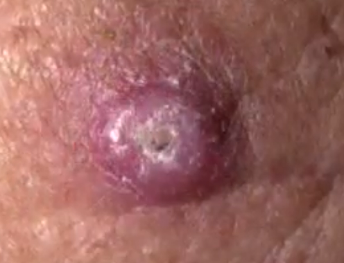
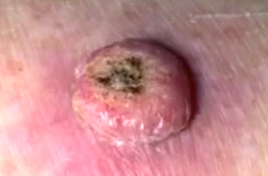
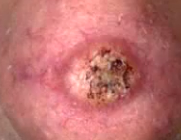
Keratoacanthoma growth is mainly found on skin that receive heavy exposure to sunlight and it affects the face, hands and forearms. The growth is characterized by dome shape, symmetrical structure and is surrounded by an inflamed skin with smooth wall. The skin is capped with debris and it has keratinized scales. One peculiar characteristic with this low-grade tumor is that it grows rapidly and can reach a large size in just a few days or weeks.
If the growth is not treated, it starves itself of nourishments, necroses or dies and sloughs to heal with a scar. Although pathologists classify Keratoacanthoma (KA) as a distinctive tumor that is not malignant, there is sufficient clinical evidence and history, which associates this tumor with squamous cell cancer. The cells can advance to cancerous stage from the low-grade tumor. When the growth appears, it needs aggressive treatment to ensure that it does not progress to cancerous form.
At first, the skin forms a red bump that is small, round and with the color of the skin. It looks like a spot but it has no pus. As the tumor grows, it assumes the shape of a small bump and becomes firm, dome-shaped and raised on the skin surface. It has a smooth surface and a plug at the center that is made up of keratin. When the plug at the center comes out, it leaves something like a mini volcano. When it heals, it leaves a puckered scar, which is flat. In most cases, the growth passes through 3 main stages which are the rapid growth stage, the static phase where it does not change and the last phase of healing.
Causes of keratoacanthoma
Keratoacanthoma could serve as a marker for Muir-Torre syndrome, which is a key autosomal dominant familial syndrome for cancer. The condition affects older people and is rare in young adults. It is also uncommon in darker skinned people and males are twice more likely to suffer than females. The exact cause for keratoacanthomas is not known but there are factors that are said to play a role in suffering the condition and they include heavy exposure to sun, getting in contact with certain chemicals.
Smoking and infection with particular strains of wart virus may also be associated with the condition. Suppressed immune system and traumas or minor injuries to the skin may also induce the condition. Similar to squamous cell cancer, studies show that ultraviolet rays from the sun can contribute to keratoacanthoma. In industrial workplace, employees exposed to pitch and tar show higher incidences of both squamous cell cancer and keratoacanthoma.
Symptoms of keratoacanthomas
This condition causes dome shaped keratinized or skin-colored bumps that look like a pimple. The tumor grows to form a nodule with depression at the center that is filled with the protein keratin. The lesions are painless although some may turn to be itchy. The keratoacanthoma can affect the normal function of the skin on the areas affected. The bumps are found on the face, back of hands, ears, scalp, forearms, and lower legs particularly in women. The growths range about 1-2.5 cm.
Keratoacanthoma Treatment
Keratoacanthoma can be treated with different techniques. When you notice a new lesion or bump on your skin in areas which are often exposed to the sun such as face, forearm, legs and hands, you may been to see a doctor for an examination. Similarly, if you get spots that bleed easily and they do not seem to heal, you also need to see a doctor or dermatologist. If you have an existing spot or lump on skin and suddenly it begins to change its color, shape, size or texture, or it begins to get itchy, bleeds and turns tender and sore, you should consult a doctor.
Although keratoacanthoma can heal on its own within 6 months, if left untreated, they could on rare incidents develop to high-grade squamous cell cancer. When keratoacanthoma disappears and resolves, it forms a depressed scar. The growths can cause damage on skin and the surrounding tissue as well as psychological stress. In yet another rare incidence, the keratoacanthoma can aggressively invade the tissue below the skin reaching the lymph glands.
A skin biopsy may be performed to determine if the growth is a keratoacanthoma. Treatment options include freezing the area with liquid nitrogen thus destroying the growth. Removal by excision may also be done using knife like instrument or the scalpel to cut the keratoacanthoma out and stitch the skin. Mohs micrographic surgery may be performed by taking tiny slivers of skin right from the affected area until the growth is removed completely especially in nose, ears and hands.
Electrodessication may be applied to scrape and burn the growing tissue. A curette is used to scrape the skin cancer cells and an electric needle is used to cauterize or burn the tissue. Radiation treatment is applied if a patient has difficulties with other treatments. A close examination after the removal of the tumor is required to monitor the skin of any possible growth recurrences.
Keratoacanthoma Pictures


