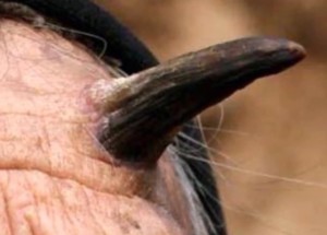
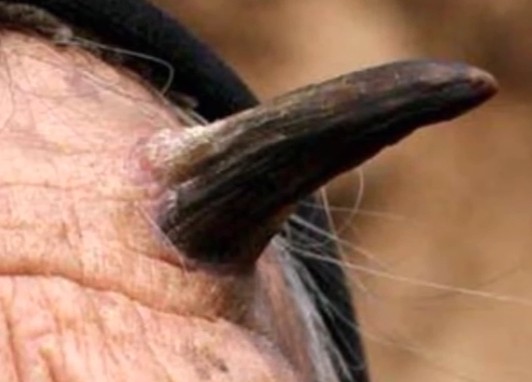
Keratin or cutaneous horn is a projectile conical shaped growth, which extends from base of skin and resembles the horn of animals. The cutaneous horn is made up of compacted keratin. Keratin is a structural material or fibrous structural protein that makes up the outer layer of skin. It is also the structural component of nails and hair, and provides the strength and toughness needed for masticatory organs such as tongue. Keratin horn is a hyperkeratotic nodule that protrudes from the skin.
Cutaneous horns mainly occur in skin areas that are exposed to burns or actinic radiation. They can be found mainly on the upper parts of face. Other locations where you may find these horns are eyelids, lip, ears, nose, scalp, neck, chest, and shoulder. The cartilaginous part of ear, back of hands, and legs may also develop these lesions.
Although 60 percent of these lesions are benign, meaning they may not signify any serious health conditions, nonetheless, there are some, which are malignant and premalignant. Some cancer conditions have been associated with keratin horns such as basal cell carcinoma, malignant melanoma, trichilemma, sebaceous molluscum, keratosis, verruca, Bowen’s disease, and epidermoid carcinoma.
In order for patients having cutaneous horn to undergo diagnosis, the lesions are studied using biopsy at base of horn. Excision may be considered in case of smaller lesions. The malignant and premalignant conditions associated with the lesions are commonly found in older males especially when the nodules are found on pinna, face, forearms, dorsum of hands, or the scalp.
The cancerous and precancerous conditions may also be found where the horn has a large base. Although cutaneous horns are asymptomatic, due to their excessive height where they protrude on skin, they could easily be traumatized. The traumatization may result in inflammation at base leading to pain.
Although the horns can arise from actinic keratosis, seborrhoeic keratosis, or viral wart, about 15 percent will form from underlying squamous cell carcinoma. Keratin horns appear firm and white to yellow in color and are often curved. In most cases, the presence of inflammation or indurations at the base of the horns is a suggestive indicator of malignant transformation to squamous cell carcinoma.
Causes of keratin horn
Different causes of cutaneous horns have been suggested. Most of the malignant lesions found at base of the horns are mainly squamous cell carcinoma. Rarely has basal cell carcinoma been reported. The malignant lesions are profoundly precipitated by UV radiation. Some rare tumors found at base are such as sebaceous adenoma, Paget disease of breast, and granular cell tumor.
A frequent finding at base of the horn is a premalignant lesion called actinic keratosis. The human papilloma virus is known to frequently cause infectious etiology, which results in a verruca vulgaris. All these conditions are associated with cutaneous horn.
Benign causes are many and they include trichilemmal cyst, seborrheic keratosis, intradermal nevus, epidermal nevus, and trichilemmoma, prurigo nodule. A trichilemmal cyst also known as wen or pilar cyst commonly forms from hair follicles. These cysts are found on scalp and they are smooth and filled with keratin.
Seborrheic keratosis is a common skin growth that might look worrisome though it is not cancer. This skin growths start as small and rough bumps and they become thick attaining a warty surface. Seborrheic keratoses growths appear white, black, tan, or brown and have a waxy look.
An intradermal nevus is a noncancerous patch of skin and is caused by overgrowth of skin cells. These are seen at birth but they may also develop in early childhood. They appear flat forming tan patches on skin that are raised. As an individual age, the epidermal nevus growths become thicker and darken taking a wart like appearance.

Symptoms and treatment
There may be no symptoms associated with cutaneous horns apart from their cone shaped and protuberance appearance, which develops out of a bump. The bump where the horn develops varies in color from red to pink or skin colored, and takes the form of a lesion that is flat. The exact cause has not been established but it is known that many of the horns will form on skin, which is exposed to sun.
It also develops on skin parts not exposed to sunlight. Some people with burn scars have also developed cutaneous horns. When the cutaneous horn is thought to be benign, there is no treatment that is needed. However, a patient needs to see a dermatologist to ensure that it isn’t cancerous or pre-cancerous.
The horns that seem to be sensitive and tender at base will mostly likely be malignant. A surgery procedure may be performed to remove the precancerous horns. Removing cancerous horns may not require surgery but use of radiation therapy. People should never attempt to remove the keratin horns by themselves because the skin may require grafting or stitches.
