Bibasilar atelectasis is a partial or complete collapsing of the lungs or lobe of lungs when alveoli, the tiny air pockets become deflated. This condition causes problems in breathing and may occur after a surgery. Atelectasis may also occur due to other problems like cystic fibrosis, lung tumor, inhalation of foreign objects, accumulation of fluid in lungs, chest injuries, and severe asthma. Usually the tissue damage varies depending on the causal factor. Atelectasis can accompany other conditions like COPD, pneumonia, lung disease and asthma. In patients who undergo surgery with general anesthesia, the condition may also manifest. This condition can be fatal since it reduces the availability of oxygen in body.
Causes of atelectasis
This condition can occur as a result of blockage in airways meaning obstructive cause. Pressure from outside the lungs meaning nonobstructive cause, may also lead to the condition. Anesthesia can cause problems in gas absorption and pressures, as well as changes in airflow in lungs. These aspects can result to collapse of the alveoli or the tiny air sacs in lungs. These conditions are often experienced after a bypass surgery.
Most people who undergo surgery have some level of atelectasis arising from the use of anesthesia substance. Another cause of atelectasis may be obstruction of the bronchial tubes or air passages. Accumulation of mucus within the bronchial tubes after a surgery can cause obstructive atelectasis. The mucus plug forms during and after surgery since a patient is not able to cough.
The drugs which are administered in a surgery procedure make the lungs to inflate less than normal, something that causes normal secretions to accumulate in the airways. Although suctioning done during surgery helps remove the secretions, they can still form after the surgery. This is why it is recommended that you breathe and cough deeply when you are recovering from surgery to help clear the secretions.
When foreign objects are inhaled in lungs as happens with children, they can induce atelectasis condition. When a person suffers from chronic infections such as tuberculosis, fungal infections and other lung conditions, they can cause scar and constrict the air passages. Tumors and blood clots are also associated with the condition.
Nonobstructive causes which can lead to atelectasis include injury in the chest, which causes chest trauma. If a person falls or is involved in a car accident, the injuries can prevent deep breathing, which in turn leads to compressed lungs. Build up of fluid between the pleura tissue in what is known as pleural effusion can cause atelectasis. Pleura are the tissues, which line the lungs and the interior of the chest wall.
Moreover, pneumonia is another cause of nonobstructive atelectasis. A large tumor outside the lungs can press against this organ causing deflation rather than blocking the airways thus causing atelectasis. Other causes are scarring of lung tissue from injury, surgery, or a lung disease.

Symptoms of atelectasis
The symptoms of atelectasis may include dyspnea or difficulties in breathing, coughing, a rapid shallow breathing, and low-grade fever. Wheezing, confusion, and coughing blood many also show up in patients suffering from this condition. In acute atelectasis, there is sudden obstruction of bronchus, cyanosis, and dyspnea as well as elevation of body temperatures. A patient may also experience shock or a drop in blood pressure.
For chronic form of the condition, the symptoms may not be noticeable but there is gradual development of dyspnea or difficulties in breathing, accompanied by body weakness. You need to see a doctor if you have problems breathing especially if you recently undergone a surgery. Other conditions can cause trouble in breathing and therefore, an accurate diagnosis is needed for the right treatment.
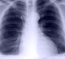
At times, there may be no apparent symptom of signs of atelectasis but the mentioned symptoms are what you need to look out for to detect if you are suffering from the condition. To diagnose the condition, doctors may use a number of procedures such as tests on complete blood count, kidney profile, examination of serum electrolytes, chest x-ray, bronchoscopy, and sputum cytology where cells of the lungs are tested of mucus for cancer.
A bronchoscopy examination is applied in the diagnosis procedure to help rule out any obstruction that may be caused by foreign body or neoplasm when the cause of atelectasis is unknown. CT scans of the chest and pulmonary function tests are other examinations that may be done to determine the condition.
Bibasilar atelectasis treatment
Treatment of this condition largely depends on the cause. When the condition is mild, it may subside without any need for treatment. However, if there are underlying causes for the condition like tumors then radiation, chemotherapy or surgery may be performed to remove the growth. A patient who undergoes surgery may get help through chest physiotherapy.
Certain techniques that are applied to help patients breathe deeply following a surgery can assist in re-expanding the collapsed lung tissue. The techniques may be learned prior to the surgery and they include coughing deeply, and clapping the chest around the collapsed tissue to loosen up the mucus. Air pulse vibrator vest may be used to mechanically clear the mucus.
Deep breathing exercises or incentive spirometry is another technique, which can help in managing the condition. Postural drainage by positioning the body with the head lower than the chest can allow the mucus to drain from the bottom of lungs. Inhaled bronchodilators can help in opening wider the bronchial tubes to make breathing easier.
Mucus thinners such as acetylcysteine can help thin mucus therefore making it easier to cough up and remove the mucus. In children who suffer from cystic fibrosis, medications like domase alfa may be used to clear mucus plugs. Supplemental oxygen may be helpful to relieve the shortness of breath caused by atelectasis. Moreover, surgical procedure may be recommended to remove obstructions in airways. Bronchoscopy is applied with use of a flexible tube, which is inserted down the throat to help clear the air passages. Suctioning also helps clear the airways of mucus.
In essence, if the atelectasis condition is acute, treatment is done by removal of the cause. The most common procedures are suctioning, coughing, and bronchoscopy. Chronic atelectasis by and large, requires use of surgical procedure to remove the part of the lube of lung, which is affected. After the surgery, antibiotics are administered to treat infection, which accompanies secondary atelectasis
Author
-
Sponsored link